Evento a cura del Centro Piattaforme Tecnologiche.
March 13th , 2017 – h. 11.00 : 13.00
Dr.ssa Giulia De Luca
Aula C Lente Didattica
Scientific Volume Imaging (SVI) Product Specialist Huygens
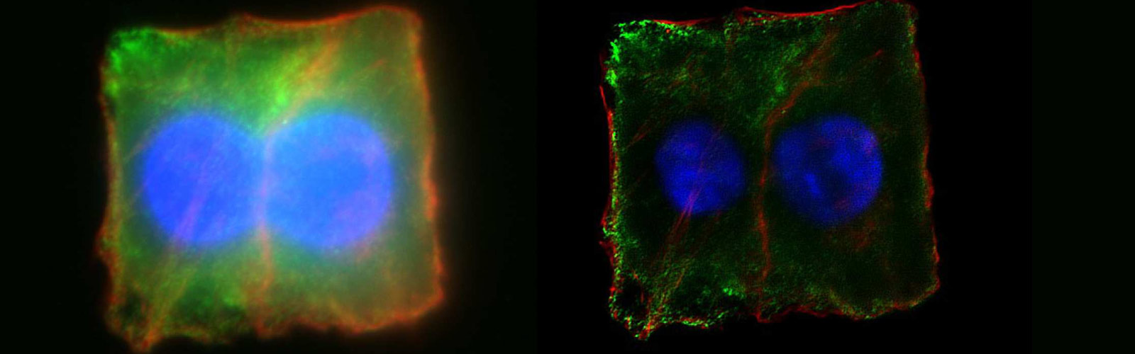
In the latest decades the study of biological processes has been strongly boosted by very powerful techniques such as wide-field fluorescence, confocal and multiphoton microscopy. However, the raw image we obtain from the sample is, by definition, an approximation, or in technical words a ‘’convolution’’ of the sample, in which the real image is spoilt by the out-offocus light, various types of noise and diffraction. The Huygens software offers high quality restoration (deconvolution), visualization and analysis for fluorescence microscopy, as confocal, widefield & brightfield, multi-photon and light sheet microscopy. Huygens represents the gold standard in the field, widely accepted by the scientific community worldwide. In this seminar the SVI Specialist Giulia De Luca will illustrate the features of the software and its application to the processing of biological sample images. After deconvolution, images will appear strikingly improved both in 3D resolution (by a factor of 2) and in contrast and signal to noise ratio (by a factor of 4 to 10). Since the process is conservative, the visualization and the localization of fluorescence after deconvolution is more precise and their analysis is more robust.
For more information visit the
www.svi.nl website.
Local organization and contact:
Dr.ssa Erika Lorenzetto: erika.lorenzetto@univr.it
Dr.ssa Maria Teresa Valenti: mariateresa.valenti@univr.it
Prof. Massimo Delledonne: massimo.delledonne@univr.it